+86-400-850-4050


Didn't find what you were looking for?
Consult a professional engineer immediately for your service


Transmission Electron Microscopy (TEM) Testing: based on the wave properties of electron beams, a high-energy electron beam emitted from an electron gun is converged by a condenser lens to penetrate the specimen. It is then sequentially magnified by an electromagnetic lens system, including objective, intermediate, and projector lenses, and finally imaged on a fluorescent screen or recorded by an imaging system.

| Project Overview
Transmission Electron Microscopy (TEM) Testing: based on the wave properties of electron beams, a high-energy electron beam emitted from an electron gun is converged by a condenser lens to penetrate the specimen. It is then sequentially magnified by an electromagnetic lens system, including objective, intermediate, and projector lenses, and finally imaged on a fluorescent screen or recorded by an imaging system.
Its remarkable advantages lie in the fact that it achieves a resolution of up to 0.05 nm, enabling the capture of atomic-level details; it offers magnification beyond one million times, thus realizing atomic-scale resolution; and, when combined with an energy dispersive spectrometer, it allows both qualitative and quantitative analysis of crystal structures, defect distributions, and elemental compositions. This technology supports research in materials science by elucidating the relationship between microstructures and properties of metals, semiconductors, and other materials; in life sciences, it enables the observation of cellular ultrastructures and viral morphology; and in physics, it provides critical experimental evidence for nanomaterials research.
Special sample preparation is required prior to testing: bulk samples are sectioned into ultra-thin slices of approximately 50 nm using an ultramicrotome; powder samples are suspended in a solvent and then dropped onto a supporting film; liquid samples are observed using a liquid cell holder equipped with a silicon nitride window.
| Test Objective
1. Observing microscopic morphology and structures: leveraging high resolution (up to 0.05 nm) and ultra-high magnification (over one million times), it allows the observation of nanoscale microstructures in materials and biological samples (such as crystal lattices, cellular organelles, and nanoparticle morphology), thereby revealing atomic-level arrangements and defect distributions.
2. Analyzing material composition and properties: in combination with attachments such as energy dispersive spectrometers, it enables qualitative and quantitative analysis of crystal structures and elemental compositions of samples, investigating the correlation between defects, dislocations, and material performance, or elucidating the spatial conformations of biological macromolecules.
3. Serving multidisciplinary research needs: in materials science, it guides the development of new materials; in life sciences, it explains disease mechanisms; and in physics, it provides experimental evidence for nanotechnology, thereby enabling correlation studies from microstructural features to macroscopic properties.
| Testing Standards
GB/T 34002-2017 Microbeam analysis-Analytical transmission electron microscopy - Methods for calibrating image magnification by using reference materials having periodic structures
GB/T 18907-2013 Microbeam analysis - Analytical electron microscopy - Selected-area electron diffraction analysis using a transmission electron microscope
JB/T 9352-1999 Test method for the transmission electron microscope
| Service Products / Fields
Transmission electron microscopy is widely applied. In the field of materials science, it enables the study of microstructures in metals, ceramics, and other materials, thereby supporting new material development. In life sciences, it facilitates the observation of cellular and viral ultrastructures, aiding research into disease mechanisms. In the semiconductor industry, it is used for observing the atomic structure inside chips and for defect analysis, improving chip production yield, while also providing support for advancements in display technology.
| Project Advantages
High resolution: up to 0.05 nm, capable of capturing atomic-level microstructural details.
Ultra-high magnification: exceeding one million times, enabling atomic-level resolution imaging.
Multifunctional analysis: can be combined with energy dispersive spectrometers and other attachments to simultaneously analyze crystal structures, elemental compositions, and defect distributions.
Cross-disciplinary applicability: meeting microscopic research needs across multiple fields, including materials science, life sciences, and semiconductors.
Non-destructive/low-damage testing: for example, cryo-electron microscopy technology can reduce beam-induced damage to biological samples.
Dynamic observation capability: certain models allow real-time monitoring of in-situ dynamic processes (such as phase transitions and material growth).
| MTT Advantages
1. Professional Team: Equipped with a number of highly experienced testing engineers and technical experts.
2. Advanced Equipment: Equipped with internationally leading testing instruments to ensure accuracy and reliability of results.
3. Efficient Service: Rapidly respond to customer needs and provide one-stop, high-efficiency inspection services.
4. Authoritative Certification: The laboratory is certified by ISO/IEC 17025, ensuring that test reports have international credibility.

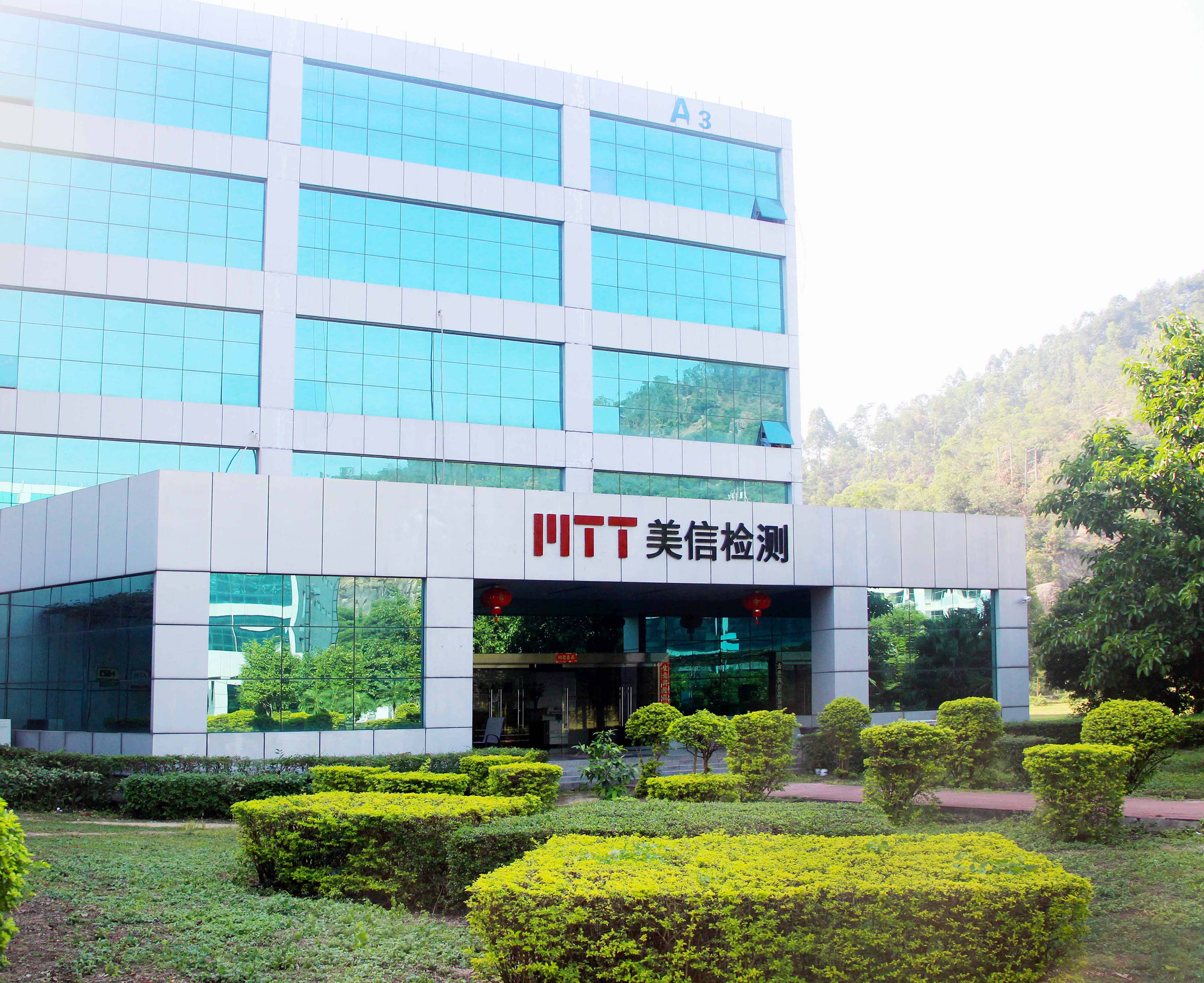
Maxin Testing Laboratory
Learn more about our company

Electronic Manufacturing Information Hub
See more exciting case studies

Video Channel
Watch more exciting videos


 Buildings A1, A3, and A5, Peking University Science and Innovation Park, Songbai Road, Bao'an District, ShenzhenBuilding 3, Xinyao Valley, 415 Changyang Street, Suzhou Industrial Park, Suzhou City1st Floor, Building 4, No. 39 Anzhi Street, Suzhou Industrial Park, Suzhou City